Author: Dr Vidisha Vaidya, Consultant Radiologist, Director of Medical Education (Clinical Radiology); RANZCR Director of Training SKG Radiology; Senior Lecturer, University of Notre Dame, Australia.

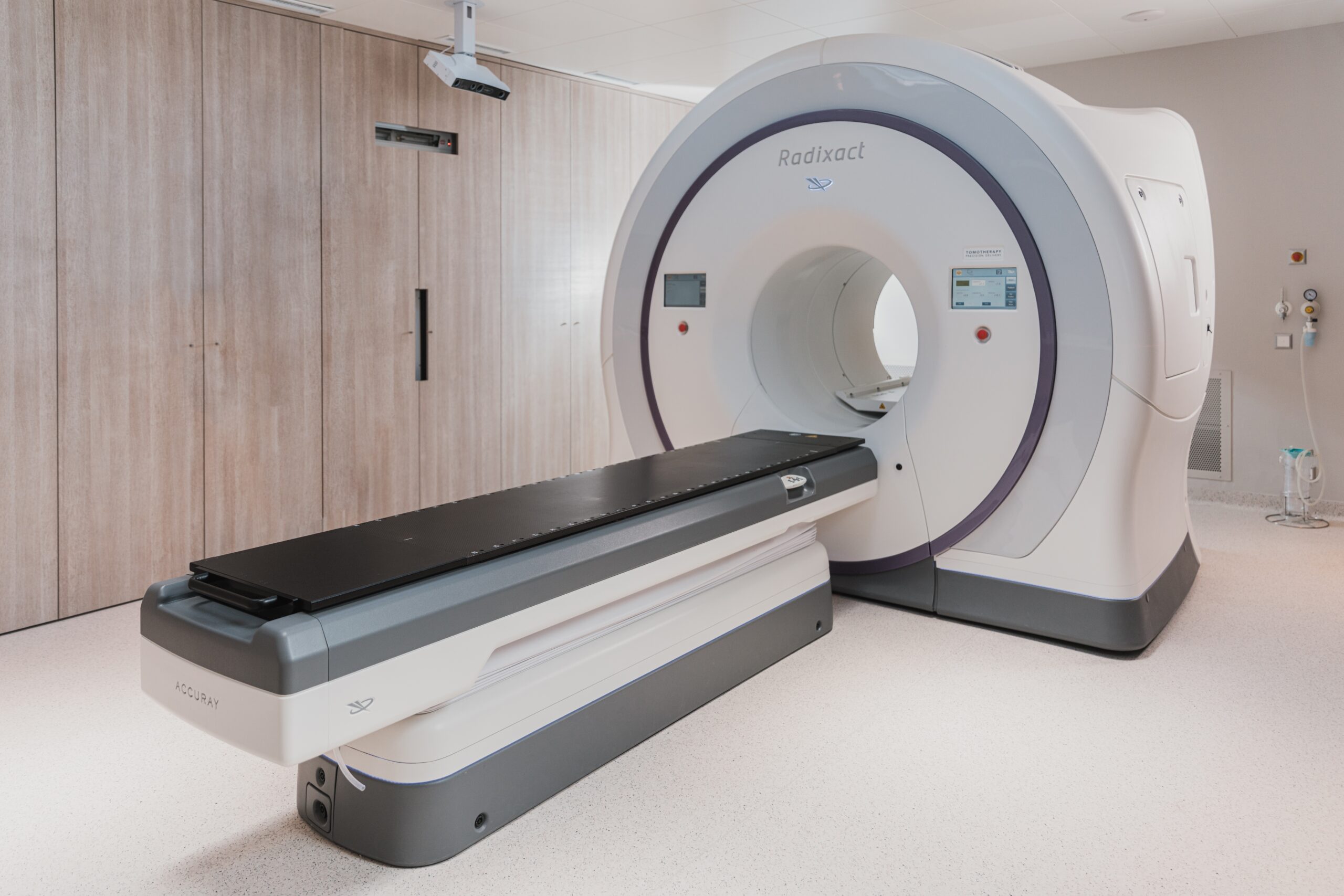
Referring and preparing patients for radiology is often performed in a sub-optimal way. Here, Dr Vidisha Vaidya, a Consultant Radiologist, takes us through her essential tips for requesting and preparing patients for CT scans.
CT scan essential considerations: 6 Cs
- Clinical question: always have a clear clinical question (e.g., exclude obstructing renal stone, confirm acute appendicitis, exclude acute intracranial haemorrhage). If unsure, ask your senior Registrar or Consultant to clarify.
- Contact details: always include your full name, designation and contact phone number (ensure contact number is not the hospital switch board number.
- Coordinate handover and specify this on the request form: if the study will be performed after your rostered working hours, always include the name and phone number of the person taking over from you. If you are unsure, look it up on the hospital intranet rosters or obtain this information from switch board and/or from the friendly ward clerk and/or nurse coordinator.
- Creatinine < 150 mmol/L and EGFR > 30 mL/min/1.73m2
- Cannula: 20 G (pink) cannula preferred. 22 G (blue) can be used for portal venous (PV) phase
- Consent: assess patient capacity. If patient is not compos mentis, consider next-of-kin consent and/or two doctor consent for IV contrast administration.
Requesting brain and head & neck CT scans
| Trauma | CT non-contrast, CT cervical spine |
| Acute stroke | Call code stroke, CT stroke protocol |
| Acute haemorrhage or known progressive haemorrhage | CT non-contrast |
| Head and neck cancer | CT contrast |
| Brain metastasis | CT non-contrast and CT contrast |
| Abscess, infection | CT with contrast |
| Dural venous sinus thrombosis | CT venogram with contrast |
Requesting intra-abdominal CT scans
| Diverticulitis, appendicitis, cholecystitis | CT abdomen, portal venous (PV) phase |
| Liver lesions (most) | CT abdomen, PV phase |
| Hepatocellular carcinoma (HCC) | CT abdomen, Tri-phasic liver |
| Acute pancreatitis | CT abdomen, PV phase (<10 days), pancreas protocol (>10 days) |
| Ischemic gut, ruptured AAA | CT abdomen, Tri-phasic CT study |
| Renal colic | CT KUB, non-contrast |


